
l’Immunofluorescenza Indiretta (IFAT o IFI) resta a tutt’oggi, insieme ad alcune metodiche ELISA (infatti non tutti i test ELISA per la ricerca degli anticorpi specifici anti-leishmania sono equivalenti) il golden standard identificato dalle linee guida in diagnostica veterinaria. Il test richiede l’utilizzo di vetrini preparati in laboratorio da colture di Leishmania spp. Solo la Di.Lab. S.r.l. tra tutti i laboratori di analisi privati in Italia ed Europa, utilizza tale procedura, che richiede personale altamente qualificato ed esperto. In questo lavoro, si pubblicano per la prima volta i diversi patterns di fluorescenza (ovvero le diverse immagini ottenute al microscopio) ottenuti utilizzando tale metodica. Già abbiamo evidenziato in un precedente lavoro che ELISA ed IFAT forniscono risultati che possono essere non confrontabili tra di loro, e che i risultati forniti dalle metodiche ELISA commerciali risentono delle scelte del produttore nello stabilire gli appropriati “cut off” per definire i campioni come positivi o negativi: infatti tali valori variano in base alle diverse zone geografiche a seconda della prevalenza della leishmaniosi. l’IFAT è invece indipendente da tale problematica, fornendo un risultato come titolo, e lasciando al medico veterinario la possibilità di interpretarlo.
ABSTRACT
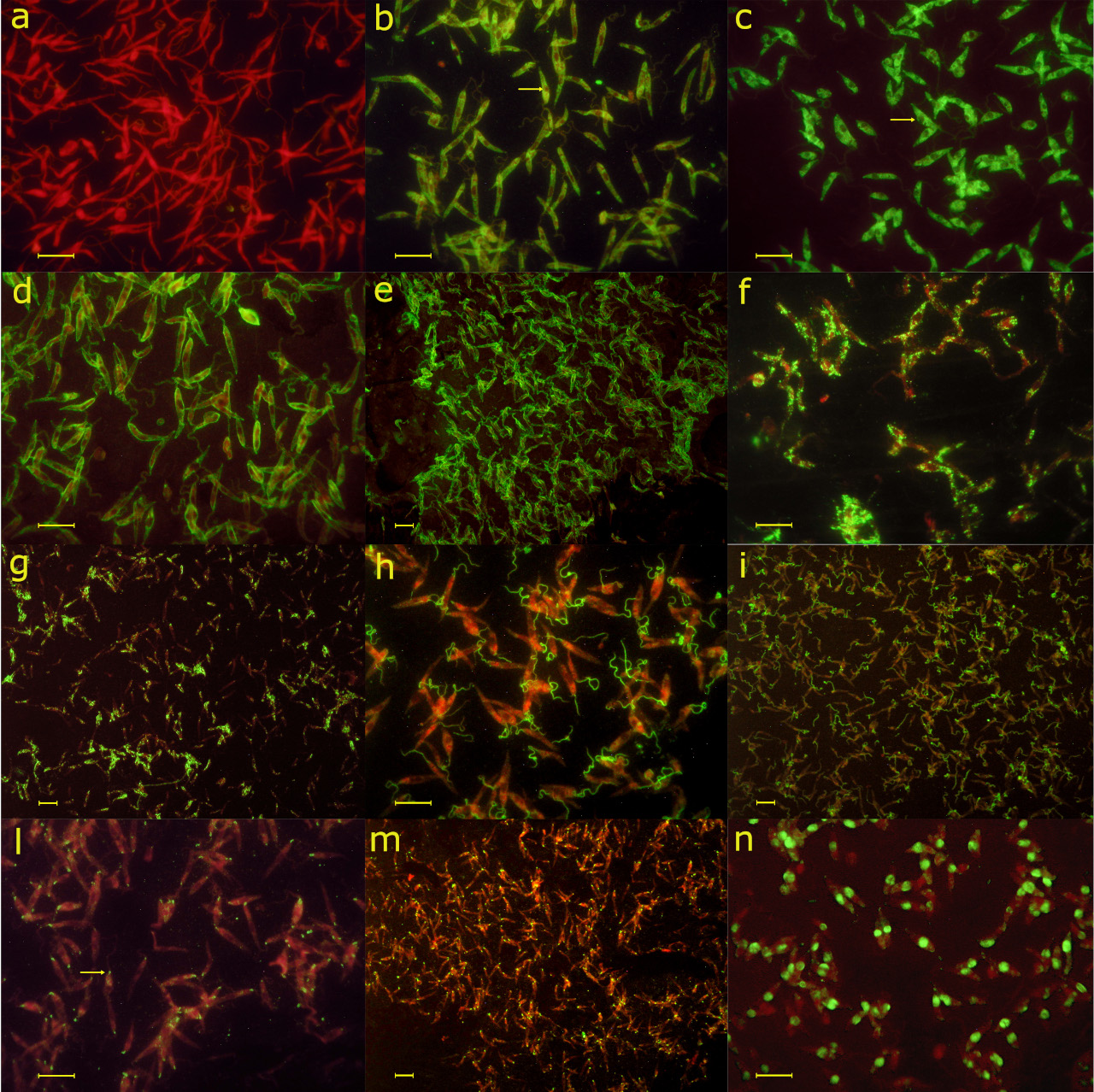
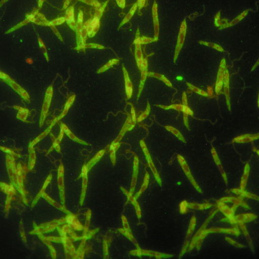
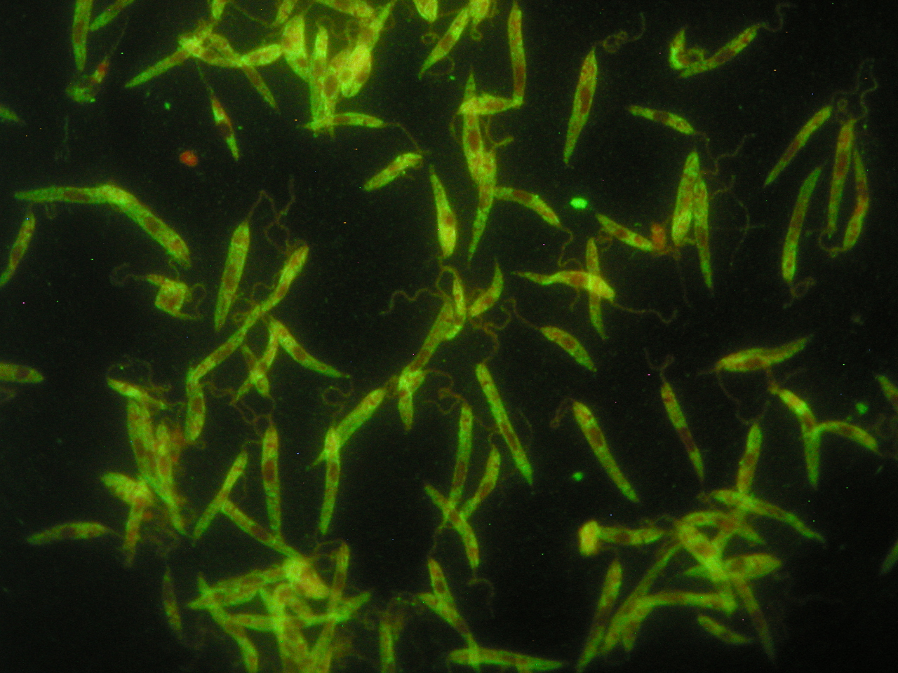
Although Indirect Immuno-Fluorescent Antibody Test (IFAT), performed employing “in house” prepared antigen, is considered by several authors as the golden standard for the quantisation of anti-leishmania antibodies in dogs, there is a lack of papers reporting a description of the different patterns of fluorescence that can be observed. An incorrect identification of patterns of fluorescence may be an important source of bias in the interpretation of results. Previous papers report different criteria to define as “positive” a specific pattern of fluorescence, namely: membrane fluorescence, homogeneous fluorescence of the body, or homogeneous fluorescence of the body plus flagellum. In this paper, we report a detailed description of preparation of slides and of the patterns of fluorescence that can be obtained employing “in house” prepared antigen. At least six main patterns of fluorescence may be observed: 1): homogeneous cytoplasmatic green fluorescence; 2): membrane pattern, in which the fluorescence is mainly localized along the entire perimeter of the parasites; 3): coarse-speckled cytoplasmatic fluorescence; 4): flagellar pattern, in which the fluorescence is localized exclusively onto the flagellum; 5): punctiform pattern, in which the fluorescence is localized exclusively at the basis of the flagellum; 6): nuclear pattern, in which only the nucleus of the parasite shows a homogeneous green fluorescent. The significance of each pattern is discussed.

